FESEM
تاريخ النشر : 0000-00-00 00:00:00
عدد المشاهدات : 161

تاريخ النشر : 0000-00-00 00:00:00
عدد المشاهدات : 161
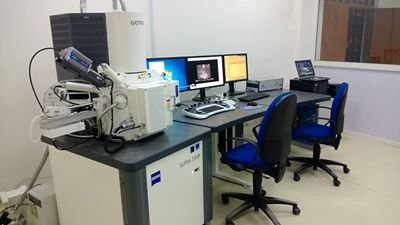
FESEM (Field-emission Scanning Electron Microscope) (SUPRA55VP)
Is a type of electron microscope that produces images of a sample by scanning it with a focused beam of electrons. The electrons interact with atoms in the sample, producing various signals that can be detected and that contain information about the sample's surface topography and composition. The electron beam is generally scanned in a raster scan pattern, and the beam's position is combined with the detected signal to produce an image. FESEM can achieve resolution better than 1 nanometer. Specimens can be observed in high vacuum, in low vacuum, in wet conditions, and at a wide range of cryogenic or elevated temperatures.
The most common mode of detection is by secondary electrons emitted by atoms excited by the electron beam. On a flat surface, the plume of secondary electrons is mostly contained by the sample, but on a tilted surface, the plume is partially exposed and more electrons are emitted. By scanning the sample and detecting the secondary electrons, an image displaying the topography of the surface is created. It enter in many applications and researchers in biology, chemistry and physics to observe small structures (as small as 1 nanometer = one billion of a millimeter!) on the surface of cells and material like organelles and nuclei of cells, synthetical polymeres and coatings of microchips.
Principles
The types of signals produced by FESEM include secondary electrons, back-scattered electrons, characteristic X-rays, light (cathodoluminescence), specimen current and transmitted electrons. Secondary electron detectors are standard equipment in all SEMs, but it is rare that a single machine would have detectors for all possible signals. The signals result from interactions of the electron beam with atoms at or near the surface of the sample. In the most common or standard detection mode, secondary electron imaging or SEI, the SEM can produce very high-resolution images of a sample surface, revealing details less than 1 nm in size. Due to the very narrow electron beam, FESEM micrographs have a large depth of field yielding a characteristic three-dimensional appearance useful for understanding the surface structure of a sample. This is exemplified by the micrograph of pollen shown above. A wide range of magnifications is possible, from about 10 times (about equivalent to that of a powerful hand-lens) to more than 500,000 times, about 250 times the magnification limit of the best light microscopes.
الجهازيعمل بطريقة النانوتكنولوجي حيث يمتاز بقابلية فحص العينات الغير موصلة بدون معاملات كما يمكن تكبير الصورة إلى أكثر من مليون مرة؛ الجهاز يحتوي على (EDS spectrophotometry) وعلى سبعة أنواع من (detectors) هي (X-ray, TEM, Coolstage, VPSE, SE1, SE2, EBSD) مما يتيح له إعطاء نتائج مشابهة للمجهر الالكتروني نوع (TEM). بالاضافة ان الجهاز يفحص تسع عينات في نفس الوقت وبنفس حالتها الطبيعية وهذا ما يميزالجهاز عن الأنواع الأخرى بإمكانية إعطاء دقة متناهية وبأقل فولتية.مثل فحص ماداخل الخلية وال(DNA)وكذلك في فحص بعض البوليمرز